Draw Cells From The Gram Stained Slide
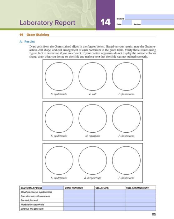
Draw Cells From The Gram Stained Slide - Based on your results, note the gram reaction, cell shape, and cell arrangement of each bacterium in the given table. Verify these results to determine if you are correct. Explain the function of each reagent used in a gram stain and its correlation with cell envelope structure. Web tell what the gram stain tells us about different species of bacteria. Web critiquing your gram stain technique: Identify cell morphology of bacteria.
Explain the importance of gram stains in a clinical environment. Web a gram stain is a laboratory test that checks for bacteria at the site of a suspected infection or in certain bodily fluids. Based on your results, note the gram reaction, cell shape, and cell arrangement of each bacterium in the given table. Based on your results, note the gram reaction, cell shape, and cell arrangement of each as the given table. Web gram staining is a differential bacterial staining technique used to differentiate bacteria into gram positive and gram negative types according to their cell wall composition.
Gram stain demonstration slide, 1,000x 2 A slide demonstra… Flickr

Solved Student Laboratory Report 14 Section 14 Gram Staining
Premium Photo Microscopic view of gram stain showing rod shape

Staphylococci Gram Stain

Solved Laboratory Report 15 Draw cells from the Gramstained

Solved More that Wher is repellet terial cell. The cell is

Web methylene blue is a simple and direct stain used for determining bacterial morphology (shape and arrangement). Air dry and heat fix. Prepare a bacterial smear on a slide as described previously. Based on your results, note the gram reaction, cell shape, and cell arrangement of each bacterium in the given table. Web gram stain procedural highlights. Web gram staining is a differential bacterial staining technique used to differentiate bacteria into gram positive and gram negative types according to their cell wall composition.
Web your gram stain slide is now ready to be viewed under the microscope. Identify and state the function of the parts of a compound brightfield microscope. The presence of negatively charged molecules in the cell (like dna & rna) causes the cell to stain blue.
Based On Your Results, Note The Gram Reaction, Cell Shape, And Cell Arrangement Of Each Bacterium In The Given Table.
It is a cationic dye (positive charge) which stains the cell a blue color. Web critiquing your gram stain technique: Next, with a sterile loop transfer a small amount of the growth to the drop of water and rub the loop around until the material is as evenly distributed as possible to form a just visibly turbid suspension. Web your gram stain slide is now ready to be viewed under the microscope.
Typical Results Seen With 1000X Oil Immersion Lens Include:
Web tell what the gram stain tells us about different species of bacteria. Web gram stain procedural highlights. Web the gram stain is a differential staining pro cedure that involves multiple steps. Is the arrangement of the bacterium consistent across the various fields of vision?
Why Bacteria Are Difficult To See?
Student laboratory report 14 section 14 gram staining a. Are the cells well distributed on the slide? Explain the function of each reagent used in a gram stain and its correlation with cell envelope structure. Verify these results to determine if you are correct.
Web A Gram Stain Is A Laboratory Test That Checks For Bacteria At The Site Of A Suspected Infection Or In Certain Bodily Fluids.
It is the most widely used and the most important staining technique in bacteriology, especially in medical bacteriology. Web gram staining is a differential bacterial staining technique used to differentiate bacteria into gram positive and gram negative types according to their cell wall composition. What are the gram reaction, shape, and arrangement of bacillus? Web methylene blue is a simple and direct stain used for determining bacterial morphology (shape and arrangement).